Elettra
BL 01.1L - TwinMic

TwinMic spectromicroscope beamline

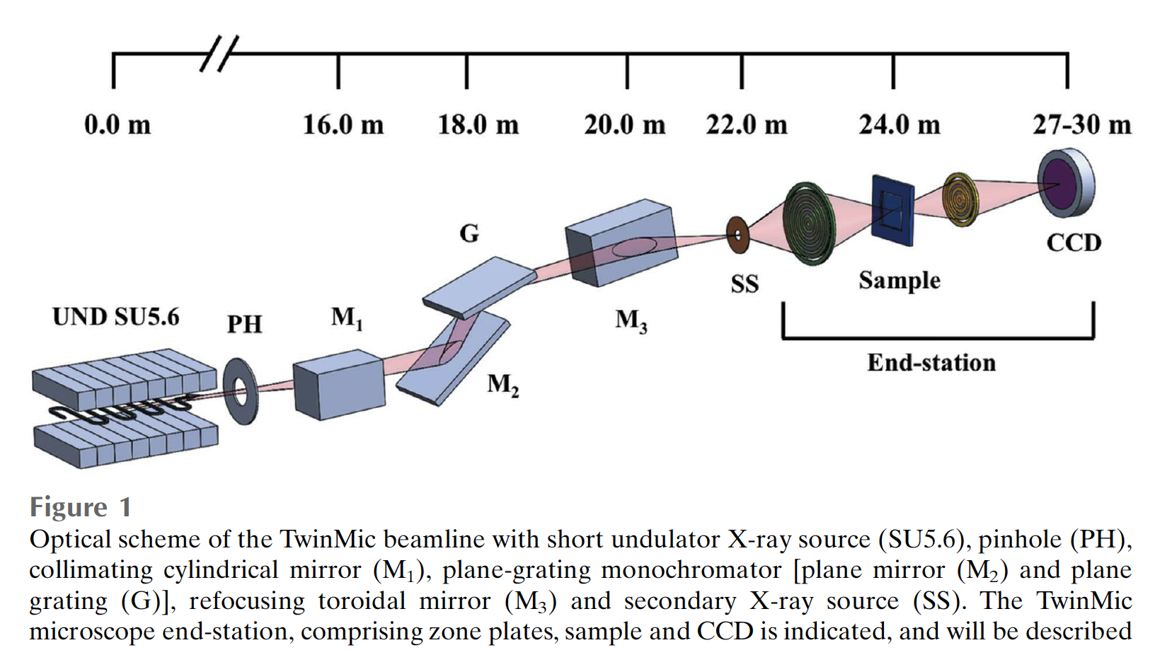
The TwinMic X-ray spectromicroscope is a world-wide unique instrument that combines full-field imaging with scanning X-ray microscope in a single instrument. The instrument is equipped with versatile contrast modes including absorption or brightfield imaging, differential phase and interference contrast or Zernike phase contrast - as you are used from a visible light microscope. The microscope is operated in the 400 - 2200 eV photon energy range or as equivalent 0.56 - 3 nm wavelengths.
According to the energy and X-ray optics TinwMic can reach sub-100nm spatial resolution.

Beamline Energy Resolution
4 * 103 [meV] @ 400 [eV]
Beamline Energy Range
400 - 2200 [eV]
Spot Size On Sample Hor
0.05 - 2 [cm]
Spot Size On Sample Vert
0.05 - 2 [cm]
Angle Of Incidence Light On Sample Value
90 [degrees]
Photon Sources

Undulator 2.0

Type
Undulator
Available Polarization
Linear horizontal
Source Divergence Sigma
X = 0.2 [urad], Y = 0.12 [urad]
Source Size Sigma
X = 700 [um], Y = 50 [um]
Energy Range
250 - 2200 [eV]
Number Of Periods
11
Period
56 [mm]

Undulator 2.4

Type
Undulator
Available Polarization
Linear horizontal
Source Divergence Sigma
X = 0.2 [urad], Y = 0.12 [urad]
Source Size Sigma
X = 700 [um], Y = 50 [um]
Energy Range
250 - 2200 [eV]
Number Of Periods
11
Period
56 [mm]
Monochromators

Monochromator 1

Energy Range
400 - 2200 [eV]
Type
PGM
Resolving Power
4000 [E/deltaE] @ 400 [eV]
Number Of Gratings
2
Grating Type
600 l/mm Au gratings
Pre-focusing Mirror Type
Cylindrical mirror
Refocusing Mirror Type
Torroidal mirror
Endstations or Setup

Endstation 1

Microscopes
- Full-field X-ray Microscope (400-800 eV) with spatial resolution down to 30nm
- Scanning X-ray microscope (400-2200 eV) provided with low energy XRF system, with spatial resolution down to 50nm at 500 eV
Spectrometer
8 Silicon Drift Detectors (SDDs) able to collect the XRF fluorescence emitted by the sample.
Photodiode available downstream the sample to acquire absorption images or absorption spectra.
Base Pressure
1 * 10-6 [mbar]
Detectors Available
EMCCD
Endstation Operative
Yes

Sample

Sample Type
Crystal, Amorphous, Other: liquid if properly sealed
Mounting Type
TEM grids, Si3N4 membranes, ultralene as supports.
For Sample Size please contact beamline personnel

Manipulator or Sample stage

Positioning Precision
X = 1 [um], Y = 1 [um]
Range Of Movement
X = 5 [mm], Y = 5 [mm]
Detectors

Silicon Drift Detector

Type
Silicon Drift Detector
Description
We have 8 SDDs for XRF imaging, 20mm2 active area
Passive or Active (Electronics)
Active

Detection

Detected Particle
Photon
Support Laboratories

sample preparation room

Description

We have a stereo visible light microscope and several basic tools such as oven, ultrasonic bath, tweezers, pipettes, mortars etc for sample preparation

contacts
Alessandra GIANONCELLI
Techniques
Absorption
  • NEXAFS
Emission or Reflection
  • Micro XRF
  • X-ray fluorescence (XRF)
Imaging
  • Fluorescence imaging
  • Medical application
  • X-ray microscopy
Address
https://www.elettra.trieste.it/elettra-beamlines/twinmic.html
control/Data analysis
Control Software Type
  • Labview
Data Output Type
  • spectra, images
Data Output Format
  • hdf5, ascii
Softwares For Data Analysis
  • PyMCA, Igor