PETRA III at DESY
P06 Hard X-ray Micro/Nano-Probe
The Hard X-ray Micro/Nano-Probe beamline P06 is dedicated to scanning X-ray microscopy with micro/nanoscopic spatial resolution using different X-ray techniques such as
- nano X-ray fluorescence (nXRF),
- nano X-ray absorption spectroscopy (nXAS),
- nano X-ray diffraction (nXRD)
- Ptychography
The beamline houses two different experiments in separate hutches. The nanoprobe experiment is optimized for ptychographic methods with resolution down to 10 nm whereas the microprobe experiment is a more versatile setup for fast scanning X-ray microscopy using KB optics providing 300 nm focus size.
Beamline Energy Resolution
1.12 [eV] @ 8000 [eV]
2 * 102 [eV] @ 8000 [eV]
2 * 102 [eV] @ 8000 [eV]
Beamline Resolving Power
1.4 * 10-4 [E/deltaE] @ 8000 [eV]
Beamline Energy Range
5 - 100 [keV]
Max Flux On Sample
4 * 1010 [ph/s] @ 10 [keV]
1 * 1012 [ph/s] @ 10 [eV]
1 * 1012 [ph/s] @ 10 [eV]
Spot Size On Sample Hor
0.08 - 1.5 [um]
Spot Size On Sample Vert
0.08 - 1.5 [um]
Divergence Hor
1 - 20 [mrad]
Divergence Vert
1 - 20 [mrad]
Photon Sources
2m U32 spectroscopy undulator
Type
Undulator
Available Polarization
Linear horizontal
Source Divergence Sigma
X = 28 [urad], Y = 4 [urad]
Source Size Sigma
X = 36 [um], Y = 6 [um]
Energy Range
3 - 100 [keV]
Number Of Periods
60
Period
31.4 [mm]
Monochromators
Cryogenic Multilayer monochromator
Energy Range
5 - 100 [keV]
Type
cryogenically cooled plane double multilayer monochromator located at 35.8 m froom the source. Plane Si X-ray mirrors (l=195 mm) are coated with three stripes: Pd/B4C (d=2.52nm), Ni/C (d=3.22 nm), Ir coating.
Resolving Power
2 * 10-2 [deltaE/E] @ 10000 [eV]
FMB OXFORD PETRA III monochromator
Energy Range
2.7 - 50 [keV]
Type
The monochromator located at 38.4 m from the source contains 2 switchable crystal optics, a Si(111) fixed exit offset crystal pair and a polished Si(111) channelcut crystal with a gap of 11.9 mm. The channelcut optic has a high energy cutoff at ~ 18 keV.
Resolving Power
1.4 * 10-4 [deltaE/E] @ 8000 [eV]
Other Optics
attenuator unit
Description
The attenuator unit is located at 87.4m from the source and provides 12 filter slides which can be used independently. The slides are equipped with Al and Si foils of various thicknesses ranging from 0.005 - 2 mm.
Horizontal deflecting flat mirror pair
Description
The beamline utilizes a pair of Silicon X-ray mirrors (length 800 mm, distance: 1000 mm) with three stripes (Chromium, Silicon and Platinum) for higher harmonic suppression. The first mirror is located at 40.8 m, 2m behind the FMB Oxford monochromator . The use of the X-ray mirrors is optional.
Prefocussing CRL
Description
Prefocusing rotationally parabolic compound refractive lenses (CRLs) made
of beryllium are located at 43.54 m from the source. The focal length of the lens system can be adapted to the
experimental needs selecting a combination of lenses from a set of 6 lens stacks (equivalent to 2x, 4x, 8x, 16x, 32x, 67.5x lenses of R = 1500 μm). It allows one to trade-off coherence properties of the beam with the intensity falling onto the entrance slits of the scanning X-ray microscope.
Endstations or Setup
Microprobe
Description
The microprobe is a versatile scanning X-ray microscope for X-ray fluorescence mapping and tomography, scanning X-ray diffraction, near edge spectroscopy and ptychography . It is located in the first experimental hutch (EH1) at 91m from the source. A powerful Kirkpatick-Baez (KB) mirror system can be used for X-ray beam focussing with spot sizes down to about 300 nm x 300 nm in air with a free working distance of 18 cm between KB optics enclosure and the focal plane. The accessible energy range is 5 - 21 keV. The sample is scannd by a three-axix piezo system (500nm travel range) on top of a air bearing rotation (for tomography applications) and large-scale stepper motors or a hexapod. An optical in-line microscope can be used for visual alignment and observation of the sample. A cryojet is optionally used for measurements under cryogenic sample conditions.
Instead of the KB system also compound refractive lenses (CRL) can be used for focussing in the extended energy range up to 35 keV (submicrometer) and 80 keV (micrometer) or for focussing the coherent part of the beam down to 100nm beam size. The microprobe stands out by the availability of the Maia energy-dispersive detector system. Inherent for the Maia system is the on-the-fly scanning mode which allows the collection of XRF spectra with sub-millisecond dwell times and thereby XRF imaging in real 3D (fluo-tomography, XANES imaging). The incremental encoder signals from the sample stages are fed directly into the Maia control unit to ensure accurate correlation between detector signal and sample position. Online data visualization is enabled using the GeoPIXE software and quantitative analysis can be performed instantly after completion of the measurement. Similar control is provided also for other detectors. Fast pixel detectors (i.e. Eiger X 4M) at the microprobe enable multimodal imaging schemes. X-ray scattering and diffraction signals can by collected synchronously with the XRF signal for single crystal or powder diffraction imaging and ptychography.
Instead of the KB system also compound refractive lenses (CRL) can be used for focussing in the extended energy range up to 35 keV (submicrometer) and 80 keV (micrometer) or for focussing the coherent part of the beam down to 100nm beam size. The microprobe stands out by the availability of the Maia energy-dispersive detector system. Inherent for the Maia system is the on-the-fly scanning mode which allows the collection of XRF spectra with sub-millisecond dwell times and thereby XRF imaging in real 3D (fluo-tomography, XANES imaging). The incremental encoder signals from the sample stages are fed directly into the Maia control unit to ensure accurate correlation between detector signal and sample position. Online data visualization is enabled using the GeoPIXE software and quantitative analysis can be performed instantly after completion of the measurement. Similar control is provided also for other detectors. Fast pixel detectors (i.e. Eiger X 4M) at the microprobe enable multimodal imaging schemes. X-ray scattering and diffraction signals can by collected synchronously with the XRF signal for single crystal or powder diffraction imaging and ptychography.
Detectors Available
Maia C 384
Vortex EM
Vortex ME4
Eiger X 4M
Vortex EM
Vortex ME4
Eiger X 4M
Endstation Operative
Yes
Sample
Sample Type
Crystal, Amorphous, Fiber
Mounting Type
diverse, please discuss with beamline staff
Nanoprobe (PtyNAMi)
Description
The PtyNAMi experiment is a scanning X-ray microscope optimized for ptychographic methods with resolution down to 10 nm. Several x-ray analytical techniques are available, such as x-ray fluorescence, diffraction/scattering, and absorption spectroscopy, giving elemental, structural, and chemical contrast. Tomographic scanning modes are routinely possible. The nanoprobe is located in the second experimental hutch (EH2) at 98m from the source. The experiment is based on nanofocusing refractive x-ray lenses (NFLs) with focus sizes down to 50 x 50 nm2 with a free working distance of several millimeter. It operated in the hard x-ray range between 10 and 30 keV. For experiments that require lower energies down to 7 keV Fresnel zone plates are used. The transverse coherence length of the beam is matched to the aperture of the focussing optics by a set of refractive x-ray lenses for prefocussing creating a secondary source. The sample is scanned by a three-axis piezo system (100nm travel range) on top of a air bearing rotation (for tomography applications) and large-scale stepper motors. An interferometric positioning system allows tracking the sample position in scanning microscopy and tomography on all relevant time scales (vibrations and long term drifts). An optical microscope can be used for visual alignment and observation of the sample. In order to exploit the various x-ray analytical contrasts, an energy dispersive silicon drift detector and various photon-counting pixel detectors for SAXS and WAXS measurements (Pilatus 300k, Lambda 750k, and in-vacuum Eiger X 4M ) are employed. While the energy dispersive detector is located on a separate table and points at the sample under 90 deg from the side, all other detectors are placed on a large detector table behind the scanner. An evacuated tube can be used to reduce the air scattering in SAXS and high-resolution WAXS geometry.
Endstation Operative
Yes
Sample
Sample Type
Crystal, Amorphous
Mounting Type
diverse, please discuss with beamline staff
Detectors
Eiger X 4M
Type
DECTRIS EIGER X 4M (vacuum compatible)
Time Resolved
Yes
Pixel Size
X = 75 [um], Y = 75 [um]
Array Size
X = 2070 [pixel], Y = 2167 [pixel]
Thickness
450 [um]
Passive or Active (Electronics)
Active
Detection
Detected Particle
Photon
Maia C 384
Type
energy dispersive Silicon X-ray detector array
Description
The Maia detector system is optimized for fast aquisition of X-ray fluorencence maps. The Maia detector array comprises 384 planar silicon 1 × 1 mm2
detector elements, 0.5 mm thick, connected to 384 independent analogue channels, each with its own charge amplification, shaping and pulse capture electronics implemented using custom Application Specific Integrated Circuits (ASICs). This strategy enables cumulative count-rates exceeding 10 M/s to be achieved with low pile-up losses.
detector elements, 0.5 mm thick, connected to 384 independent analogue channels, each with its own charge amplification, shaping and pulse capture electronics implemented using custom Application Specific Integrated Circuits (ASICs). This strategy enables cumulative count-rates exceeding 10 M/s to be achieved with low pile-up losses.
Pixel Size
X = 1000 [um], Y = 1000 [um]
Array Size
X = 20 [pixel], Y = 20 [pixel]
Thickness
500 [um]
Passive or Active (Electronics)
Active
Detection
Detected Particle
Photon
Vortex EM
Type
Energy dispersive Silicon drift detector
Description
Single element detector with 50 mm2 active area. Energy resolution ~ 160 ev@5.9 keV. Combined with Xpress3 signal processor.
Thickness
350 [um]
Passive or Active (Electronics)
Active
Detection
Detected Particle
Photon
Vortex ME4
Type
Energy dispersive Silicon drift detector
Description
Four element detector with 50 mm2 active area each. Energy resolution ~ 160 ev@5.9 keV. Combined with Xpress3 signal processor.
Thickness
350 [um]
Passive or Active (Electronics)
Active
Detection
Detected Particle
Photon
Techniques
Absorption
- NEXAFS
Diffraction
- Crystallography
- Powder diffraction
Emission or Reflection
- Micro XRF
Imaging
- Coherent diffractive imaging
- Fluorescence imaging
- Medical application
- Ptychography
- X-ray microscopy
- X-ray tomography
Scattering
- Coherent scattering
Disciplines
Chemistry
- Catalysis
- Electrochemistry
- Physical Chemistry
Earth Sciences & Environment
- Mineralogy
- Natural disaster, Desertification & Pollution
- Plant science
- Technique Development - Earth Sciences & Environment
Energy
- Sustainable energy systems
Humanities
- Cultural Heritage
Life Sciences & Biotech
- Food quality and safety
- Medicine
- Molecular and cellular biology
Material Sciences
- Knowledge based multifunctional materials
- Metallurgy
Physics
- Hard condensed matter - structures
- Nanophysics & physics of confined matter
- Optics
Address
Beamline P06 at PETRA III
Deutsches Elektronen-Synchrotron DESY
Max von Laue Hall / Geb 47c EG Sektor 4
Notkestraße 85
22607 Hamburg
Germany
Deutsches Elektronen-Synchrotron DESY
Max von Laue Hall / Geb 47c EG Sektor 4
Notkestraße 85
22607 Hamburg
Germany
control/Data analysis
Control Software Type
- Sardana, TANGO, Python
Data Output Type
- spectra, images
Data Output Format
- hdf5
Softwares For Data Analysis
- PyMCA, GeoPixe
Equipment That Can Be Brought By The User
sample holders, flow reactors, diamond anvil cells, etc. Please contact beamline staff.
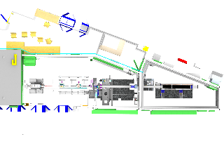
Layout
