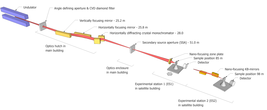
The hard X-Ray nanoprobe of Max IV, NanoMAX, is designed to take full advantage of MAX IV’s exceptionally low emittance and the resulting coherence properties of the X-ray beam. The use of diffraction-limited optics will allow producing tightly focused coherent beams enabling imaging applications using diffraction, scattering, fluorescence and other methods, at unprecedented resolution. With its two experimental stations designed to offer, the first, the smallest focal spot, and the other, large flexibility at the expense of a larger focal spot for various scattering geometries, NanoMAX will offer exciting applications for a wide variety of research fields, such as materials science, life science, earth science, nanoscience, physics, chemistry and biology.
KB experimental station
The endstation is based on a goniometer setup mounted in stable granite foundation.
A two-circle goniometer holds a coarse sample scanner (20 mm stroke).
The sample is mounted on a fine sample scanner (100 μm stroke in all directions) on the coarse scanner.
Max res: 5 μm
Max Field of view: 0.3 - 4 mm
3-element Ge detector
XSPRESS3 digital pulse processor
Sample
TEM grid.
Manipulator or Sample stage
Theta: stroke [-5, 90] deg resolution: 10-4 deg
Phi: stroke [-180, 180] deg resolution: 10-4 deg
Theta is horizontal and perpendicular to the X-ray beam. Phi is perpendicular to Theta and vertical for Theta=0.
In-house development.
Option 1 (standard), NPXY100Z100-135 (This option provides the stiffest frame and the highest resolution).
100 μm stroke in all directions.
Option 2, NPXY200Z100-271.
200 μm stroke in each horizontal direction, 100 μm vertical stroke
Sample Environment
This system was supplied by Alemnis AG, and is used for in-situ nano-XRD.
Maximum load is 0.5 N.
The system is made available in collaboration with the Sample Environment and Detector Support team at MAX IV.
Sample Holders
3.2 mm diameter mounting pin.
4 x 4 mm2 overall plate size.
2.7 mm diameter sample hole in plate.
Multi-sample holder plate.
3 slots, 3 x 5 mm2.
2 holes, 3.2 mm diameter.
12.7 mm diameter with 3.2 mm pins.
Crytur in-line detector
Detection
Diffraction detector
Detection
Eiger 4M in-line detector
Detection
Ge X-ray fluorescence detector
Detection
Pilatus 100k in-line detector
Detection
Pilatus 1M in-line detector
Detection
SDD X-ray fluorescence detector
Detection
- Fluorescence imaging
- Coherent scattering
- Elastic scattering
- Other - Chemistry
- Other - Life Sciences & Biotech
- Other - Material Sciences
Fotongatan 2
224 84 Lund
Sweden
- TBC
- TBC
- TBC